
Precise Wound Measurement: The Foundation of Effective Care
Accurate wound measurement is crucial for effective wound management and improved patient outcomes. This listicle outlines seven key wound measurement techniques, from basic ruler measurements to advanced 3D scanning. Learning the strengths and limitations of each method helps healthcare providers and patients select the best approach for accurate assessment, treatment decisions, and healing progress tracking. This knowledge empowers better wound care for all. This article covers ruler/manual measurement, digital planimetry, 3D structured light wound scanning, stereophotogrammetry, laser-assisted wound measurement, acetate tracing/contact planimetry, and mobile application-based wound measurement.
1. Ruler/Manual Measurement
Ruler/manual measurement is the most fundamental and commonly used technique for assessing wound dimensions. This method employs a simple, disposable ruler or measuring tape to determine the wound’s length, width, and often its depth. Clinicians typically measure the longest length of the wound (head-to-toe) and the widest width (perpendicular to the length). These measurements are then used to calculate the wound area using the formula: length × width. This provides a quantifiable assessment of the wound size, which is crucial for monitoring healing progress and determining the effectiveness of treatment.
This technique deserves its place on the list of wound measurement techniques due to its widespread accessibility and simplicity. It’s a cornerstone of wound assessment in various healthcare settings, from primary care offices to specialized wound care clinics. Key features include the use of readily available disposable rulers or measuring tapes, measurements documented in centimeters, and the frequent inclusion of depth measurement using a cotton-tipped applicator. For more complex wound shapes, clinicians may also incorporate wound tracing onto transparent film to create a more accurate representation. You can learn more about Ruler/Manual Measurement to understand the nuances of this fundamental technique.
Pros:
- Cost-effective: Requires minimal equipment, making it an economical option for all healthcare settings.
- Accessibility: Available in virtually all clinical environments.
- Ease of use: Requires no specialized training or technology.
- Speed: Quick to perform, providing immediate results.
Cons:
- Inter-rater reliability: Different clinicians may obtain slightly different measurements, impacting consistency.
- Oversimplification: Calculates area based on a rectangular shape, which may not accurately reflect irregular wounds.
- Limited sensitivity: May not capture subtle changes in wound morphology.
- Cross-contamination risk: Reusing rulers poses a risk of infection transmission.
- Human error: Susceptible to errors in measurement and documentation.
Examples of Implementation:
Ruler/manual measurement is standard practice in most wound care clinics, hospitals, and home health settings. The Bates-Jensen Wound Assessment Tool, a widely used instrument for comprehensive wound assessment, incorporates manual measurement as a key component.
Tips for Accurate Measurement:
- Always use a disposable ruler to prevent cross-contamination.
- Measure the length from head-to-toe and the width from side-to-side, maintaining consistent orientation regardless of the wound’s position.
- Ensure consistent patient positioning during serial measurements to minimize variations.
- Thoroughly document the measurement technique used to ensure consistency across different assessments and clinicians.
- Consider using wound tracing for irregular wounds to obtain a more precise representation of the wound area and perimeter.
When and Why to Use This Approach:
Ruler/manual measurement is appropriate for initial and ongoing wound assessments in most clinical situations. It’s particularly useful for quickly obtaining baseline measurements and tracking gross changes in wound size. While it may not be the most precise method for complex or irregularly shaped wounds, its simplicity and accessibility make it a valuable tool for a wide range of healthcare professionals caring for patients with acute or chronic wounds. This includes primary care providers, long-term care staff, wound care specialists, and even patients and caregivers in home settings. This simple method provides valuable information for Medicare and insurance beneficiaries, particularly elderly patients, who often require ongoing wound care and monitoring.
2. Digital Planimetry
Digital planimetry represents a significant advancement in wound measurement techniques, offering a more precise and objective approach compared to traditional methods. This technique leverages digital imaging and software analysis to accurately calculate wound area. The process involves capturing a digital photograph of the wound alongside a known calibration marker, such as a ruler. Specialized software then utilizes this marker to establish a scale and traces the wound perimeter, allowing it to calculate the precise surface area regardless of the wound’s shape. This makes it particularly useful for irregularly shaped wounds, where manual measurements can be less accurate.
Digital planimetry systems offer several advantages. Some can measure not only area but also wound volume and depth, providing a more comprehensive understanding of wound characteristics. The digital format allows for permanent visual documentation, facilitating longitudinal tracking of wound healing progress and enabling healthcare providers to detect even subtle changes in wound size over time. This digital record also reduces subjectivity in measurements, promoting consistency across different clinicians. Moreover, because it’s a non-contact method, digital planimetry minimizes the risk of infection compared to methods involving direct contact with the wound. You can learn more about Digital Planimetry to understand the full scope of its capabilities.
Examples of commercially available digital planimetry systems include VISITRAK™ by Smith & Nephew, PictZar® Digital Planimetry Software by BioVisual Technologies, and WoundVision Scout™. These systems have been instrumental in popularizing the technique and demonstrating its efficacy in various wound care settings.
While digital planimetry offers many benefits, it also has some limitations. It requires access to a digital camera or smartphone and specialized software, which can be expensive. Accurate measurements depend on consistent lighting, camera angle, and distance between the camera and the wound, which can require some practice to master. There is also a learning curve associated with operating the software, although most systems are designed to be user-friendly. Finally, the process can be slightly more time-consuming compared to a simple ruler measurement, especially initially.
Tips for Effective Digital Planimetry:
- Consistent Lighting: Maintain consistent lighting conditions for serial assessments to ensure accurate comparisons over time. Avoid shadows and reflections.
- Calibration Marker Placement: Position the calibration marker in the same plane as the wound, ensuring it’s clearly visible and adjacent to the wound.
- Perpendicular Photography: Take photographs from directly above the wound (90-degree angle) to minimize distortion and ensure accurate area calculation.
- Complete Visibility: Ensure the entire wound and calibration marker are visible within the camera frame.
- Color Calibration: Utilize color calibration features when available for more accurate tissue analysis and wound assessment.
Digital planimetry is particularly valuable in situations where accurate and consistent wound measurement is crucial, such as in clinical trials, research studies, and for patients with complex or chronic wounds. Its ability to detect subtle changes and provide objective data makes it an important tool for monitoring wound healing progress and informing treatment decisions. For patients, primary care providers, long-term care facilities, wound care specialists, and insurance beneficiaries dealing with chronic or acute wounds, particularly in elderly patients, digital planimetry offers a valuable tool for enhancing the accuracy and effectiveness of wound care.
3. 3D Structured Light Wound Scanning
3D structured light wound scanning represents a significant advancement in wound measurement techniques, offering a highly accurate and non-contact method for assessing even the most complex wounds. This technique deserves its place on this list due to its ability to provide precise measurements of volume, depth, and surface area, going beyond the capabilities of traditional methods. This makes it an invaluable tool for tracking healing progress and making informed treatment decisions.
This innovative approach works by projecting patterns of structured light onto the wound surface. Multiple cameras then capture the deformation of these light patterns as they fall on the wound bed. Sophisticated software algorithms analyze the captured data to construct a detailed 3D model of the wound, providing a comprehensive topographical map. This allows clinicians to visualize the wound in three dimensions and obtain accurate measurements of its volume, depth, surface area, and other key parameters.
Features and Benefits:
- Projects structured light patterns: This creates the basis for the 3D reconstruction.
- Uses multiple cameras: Enables capture of data from different angles for a complete 3D model.
- Creates detailed topographical maps: Provides a visual representation of the wound bed’s contours.
- Provides accurate volume and depth measurements: Offers true volume calculation, not just an estimation.
- Outputs computerized 3D models: Allows for digital storage, analysis, and comparison over time.
Pros:
- Highly accurate measurement of complex wound geometries: Ideal for wounds with irregular shapes, undermining, or tunneling.
- Provides true volume calculation (not estimated): Offers more precise data for monitoring wound healing.
- Non-contact method eliminates risk of contamination: Enhances patient safety and comfort.
- Can detect undermining and tunneling: Provides critical information not easily obtained with traditional methods.
- Excellent for tracking healing progress of complex wounds: Offers quantifiable data for objective assessment.
Cons:
- Expensive specialized equipment required: Can be a barrier to widespread adoption.
- May require technical expertise to operate: Training is necessary for accurate and reliable measurements.
- Some systems are bulky and not portable: Limits usability in certain settings.
- Initial setup and calibration can be time-consuming: Impacts workflow efficiency.
- Not widely available in all clinical settings: Access may be limited depending on location and resources.
Examples of 3D Structured Light Wound Scanners:
- eKare inSight® 3D wound measurement system
- ARANZ Medical’s Silhouette® system
- WoundVision’s Scout™ 3D camera
Tips for Using 3D Structured Light Wound Scanning:
- Ensure proper calibration before each use for accurate measurements.
- Position the scanner at the recommended distance from the wound, as specified by the manufacturer.
- Scan from multiple angles, especially for complex wounds with undermining or tunneling, to capture complete 3D data.
- Clean the optical components regularly according to manufacturer guidelines to maintain optimal image quality.
- Use wound positioning aids to ensure reproducible patient positioning and consistent measurements over time.
When and Why to Use This Approach:
3D structured light wound scanning is particularly beneficial for patients with chronic or complex wounds, such as pressure ulcers, diabetic foot ulcers, and surgical wounds. It provides valuable information for clinicians in various settings, including primary care, long-term care facilities, and specialized wound care clinics. The accurate and detailed measurements provided by this technique are crucial for:
- Monitoring wound healing progress: Objective data allows for effective evaluation of treatment efficacy.
- Making informed treatment decisions: Precise measurements aid in selecting appropriate interventions.
- Improving patient outcomes: Accurate assessment contributes to better wound management and faster healing.
This technology represents a significant step forward in wound care, offering precision and detail that can greatly benefit both patients and healthcare providers. While the cost and technical requirements may present challenges, the advantages of 3D structured light wound scanning make it a valuable investment for improving wound assessment and management.
4. Stereophotogrammetry
Stereophotogrammetry represents a significant advancement in wound measurement techniques, offering a non-contact, three-dimensional approach to assess wound healing progress. This technique utilizes two or more photographs taken from different angles to create a 3D model of the wound. By triangulating points visible in multiple images, stereophotogrammetry can accurately determine wound dimensions, surface area, and crucially, volume – a measurement often difficult to obtain with traditional 2D methods. This comprehensive data provides clinicians with a more complete understanding of wound characteristics and allows for more precise monitoring of healing over time, making it a valuable tool in wound management.
This method works by applying photogrammetric principles. Specialized software analyzes the parallax differences – the apparent shift in an object’s position when viewed from different angles – between the captured images. This analysis allows the software to reconstruct the wound’s 3D structure, including its depth and contours. Moreover, some advanced stereophotogrammetry systems can also analyze color and texture, providing further insights into the wound bed’s condition. This detailed information is crucial for accurate diagnosis, treatment planning, and monitoring the effectiveness of interventions.
Examples of successful implementation include:
- Wound3D system: A dedicated stereophotogrammetry system designed specifically for wound assessment.
- 3DWoundView: Another example of commercially available software utilizing this technology.
- Implementation in various university hospital wound care centers: Leading medical institutions have adopted stereophotogrammetry as a standard practice for precise wound measurement.
When and Why to Use Stereophotogrammetry:
This technique is particularly useful for complex wounds or those with irregular shapes where accurate volume assessment is crucial. It’s especially beneficial for chronic wounds, burns, and ulcers where precise monitoring is essential for effective treatment. The non-contact nature of stereophotogrammetry is also advantageous for fragile wounds or those at high risk of infection, as it eliminates the need for direct contact with the wound bed.
Pros:
- More accurate than 2D photography for volume assessment.
- Can be implemented with standard digital cameras, potentially reducing costs compared to specialized equipment.
- Non-contact method prevents wound bed disturbance and minimizes infection risk.
- Provides permanent visual documentation for detailed record-keeping and analysis.
- Can detect subtle changes in wound healing over time, enabling early intervention if needed.
Cons:
- Requires precise camera positioning and calibration to ensure accurate measurements.
- Complex wounds with undermining or deep tracts may be challenging to measure accurately.
- Specialized software is required for analysis, which can involve a learning curve for healthcare professionals.
- More time-consuming than simple photography, potentially impacting workflow.
- Results depend on image quality and consistent positioning, requiring careful technique.
Tips for Effective Stereophotogrammetry:
- Use a tripod or positioning device for consistent camera placement between imaging sessions.
- Include calibration markers in each image to ensure accurate scaling and measurements.
- Maintain consistent lighting between sequential assessments to minimize variations in image quality.
- Take images from at least three different angles for best results, providing more data for 3D reconstruction.
- Ensure the entire wound is visible in all photographs to capture a complete representation of its dimensions.
Stereophotogrammetry earns its place among essential wound measurement techniques due to its ability to provide accurate 3D information, particularly volume, without disturbing the wound bed. While it requires specialized software and careful technique, the benefits of precise measurement and non-contact assessment make it a valuable tool for optimizing wound care and improving patient outcomes. This technique holds significant promise for advancing wound care, especially for patients with chronic or complex wounds where precise monitoring is paramount.
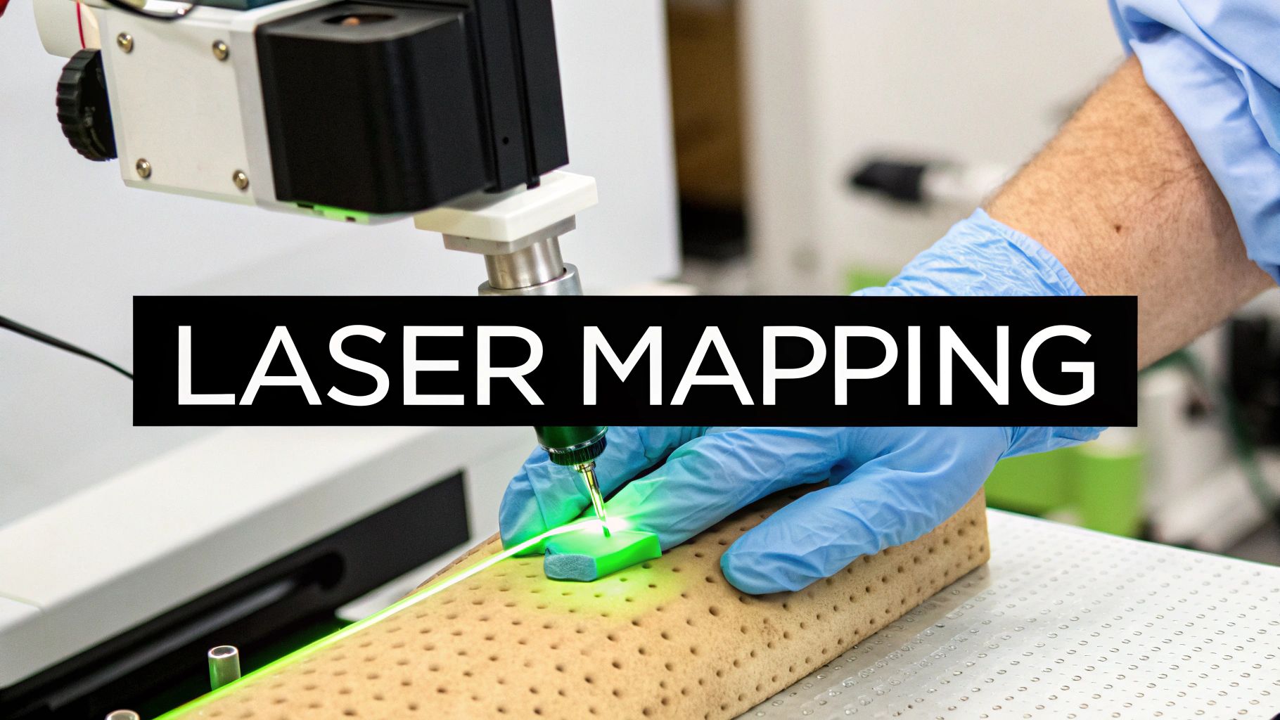
5. Laser-Assisted Wound Measurement
Laser-assisted wound measurement represents a significant advancement in wound assessment technology, offering unparalleled precision and detail. This non-contact wound measurement technique utilizes laser light to create a precise three-dimensional map of the wound, providing highly accurate measurements of area, depth, and volume. This makes it a powerful tool in various healthcare settings for accurately monitoring wound healing progress and making informed treatment decisions. This sophisticated approach stands out among other wound measurement techniques due to its ability to capture minute changes and complex wound geometries.
How it Works:
The technology works on the principle of laser displacement. A laser beam is projected onto the wound surface, and the time it takes for the light to reflect back to the sensor is measured. This time-of-flight measurement is then used to calculate the distance between the sensor and the wound surface at numerous points, creating a detailed topographical map. Some advanced systems combine laser scanning with digital imaging, providing both precise measurements and visual documentation of the wound.
Features and Benefits:
- High Precision: Laser-assisted systems offer micron-level precision, capturing even the smallest changes in wound dimensions. This level of detail is crucial for accurately tracking healing progress, especially in slow-healing or complex wounds.
- Non-Contact: The non-contact nature of this technique eliminates the risk of contamination or further injury to the wound bed, promoting patient comfort and minimizing the potential for infection.
- Topographical Mapping: Unlike traditional methods, laser scanning creates a detailed 3D map of the wound, revealing crucial information about its depth, volume, and undermining, which can be difficult to assess with other techniques.
- Reduced Interobserver Variability: The automated nature of laser measurement significantly reduces the variability between different clinicians, providing more consistent and objective data for accurate assessment and comparison.
Pros and Cons:
Pros:
- Extremely high measurement precision
- Completely non-contact methodology
- Ideal for tracking small changes in healing wounds
- Can measure difficult wound geometries including undermining
- Reduces interobserver variability
Cons:
- Specialized and expensive equipment
- May require technical training to operate correctly
- Some systems are not portable
- May be affected by ambient lighting conditions
- Limited availability in clinical settings
Examples of Successful Implementation:
- Silhouette® Star by ARANZ Medical: This commercially available system is widely used for wound assessment and research.
- FastScan™ laser scanning systems: These systems, adapted for wound care, demonstrate the versatility of laser technology in this field.
- Research Implementations at Major Burn Centers: Leading burn centers often utilize laser-assisted wound measurement for precise monitoring of burn wounds and research into novel treatment strategies.
Tips for Effective Use:
- Calibrate the system before each measurement session to ensure accuracy.
- Dim ambient lighting to improve laser visibility and measurement precision.
- Use positioning aids to ensure consistent patient positioning between measurements.
- Follow the manufacturer’s recommendations for scanning distance and other operating parameters.
- Consider using complementary color imaging for a comprehensive tissue assessment alongside the 3D measurements.
When and Why to Use This Approach:
Laser-assisted wound measurement is particularly valuable in situations where high precision and detailed topographical information are crucial:
- Chronic Wounds: Tracking subtle changes in chronic wounds can help assess the effectiveness of treatment interventions.
- Complex Wounds: Wounds with irregular shapes, undermining, or tunneling benefit from the 3D mapping capabilities of laser systems.
- Research Settings: The high precision and objectivity of laser measurement make it ideal for clinical trials and research studies.
- Burn Wounds: Accurate assessment of burn depth and area is crucial for appropriate treatment planning.
Popularized By:
ARANZ Medical, Polhemus (FastScan), and wound healing research departments at major medical centers have been instrumental in developing and popularizing laser-assisted wound measurement techniques. While this technology continues to evolve and become more accessible, it already plays a vital role in advancing wound care practices. It deserves its place among wound measurement techniques as the gold standard for precision and comprehensive wound assessment.
6. Acetate Tracing/Contact Planimetry
Acetate tracing, also known as contact planimetry, is a wound measurement technique that offers a balance between simplicity and accuracy, making it a valuable tool in various healthcare settings. This method involves placing a transparent, sterile acetate sheet directly over the wound bed and tracing the wound’s perimeter with a permanent marker. This creates a physical template of the wound’s shape, capturing its intricacies more effectively than simple length-width measurements. The tracing can then be analyzed to determine the wound’s surface area through various methods, ranging from manually counting squares on a pre-printed grid to utilizing digital scanning and software analysis. This versatility allows for adaptation to different resource levels and technological capabilities.
Acetate tracing earns its place among essential wound measurement techniques due to its ability to accurately assess irregularly shaped wounds, a common characteristic in chronic conditions. It provides a more precise measurement compared to linear methods, leading to better tracking of wound healing progress and more informed treatment decisions. Furthermore, the method allows clinicians to trace undermining and tunneling, providing a comprehensive understanding of the wound’s three-dimensional characteristics. Often, wound depth is also measured using a probe in conjunction with the tracing, further enhancing the assessment.
Features and Benefits:
- Uses transparent, sterile acetate sheets: Ensures clear visibility of the wound and minimizes contamination risk.
- Direct tracing of wound margins: Captures the precise shape and irregularities of the wound.
- Can be combined with grid counting or digital analysis: Offers flexibility in analysis methods depending on available resources.
- Creates a physical template of the wound: Enables storage in patient charts for longitudinal monitoring.
- Often includes wound depth measurement: Provides a more complete picture of the wound’s characteristics.
Pros:
- More accurate than simple length-width measurements for irregular wounds.
- Creates a physical record that can be stored in patient charts.
- Relatively inexpensive compared to digital methods.
- Minimal technology requirements.
- Can trace undermining and tunneling.
Cons:
- Contact with wound surface creates a contamination risk.
- Pressure applied during tracing may distort wound margins.
- Time-consuming process.
- Potential discomfort for patients.
- Accuracy depends on the tracer’s skill.
Examples:
- VISITRAK™ acetate sheets from Smith & Nephew offer a standardized approach to this technique. (Unfortunately, a direct link to this specific product is not readily available on their website.)
- Acetate tracing is a common practice in many wound care clinics, particularly those with limited access to advanced digital technologies.
- The VISITRAK™ system also offers a digitizer that can be used with the acetate tracings, creating a hybrid approach that combines the benefits of physical tracing with digital analysis.
Tips for Effective Acetate Tracing:
- Use only sterile acetate sheets to prevent contamination.
- Apply minimal pressure when tracing to avoid distorting the wound margins.
- Mark anatomical landmarks on the tracing for consistent orientation and comparison over time.
- Use disposable measuring devices/probes with each patient to prevent cross-contamination.
- Consider pre-printed grid acetate sheets for easier area calculation, especially in settings without access to digital scanners.
When and Why to Use Acetate Tracing:
This wound measurement technique is particularly useful for patients with chronic wounds, such as pressure ulcers, diabetic ulcers, or venous leg ulcers, where accurate assessment of irregular shapes and changes over time is crucial. It is a valuable tool for primary care providers, long-term care facilities, wound care specialists, and other healthcare practitioners involved in wound management. This technique can also be helpful in documenting wound progression for insurance purposes and demonstrating the effectiveness of interventions to Medicare and other beneficiaries. While digital methods offer advanced features, acetate tracing remains a relevant and accessible option, especially for those seeking a cost-effective and reliable wound measurement technique.
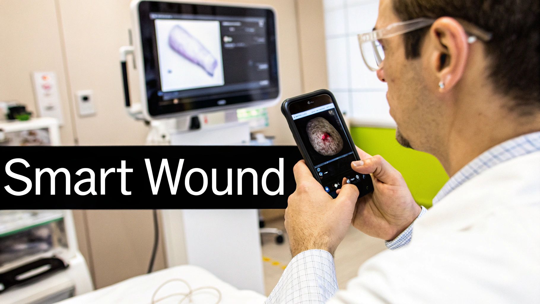
7. Mobile Application-Based Wound Measurement
Mobile application-based wound measurement is a rapidly evolving technique leveraging the ubiquity of smartphones and tablets to assess and monitor wounds. This method uses the device’s camera and specialized software to capture wound images and calculate dimensions, offering a portable, cost-effective alternative to traditional wound measurement techniques. This deserves a place on this list because it represents a significant advancement in accessible and convenient wound care, particularly for remote monitoring and telemedicine applications. Learn more about Mobile Application-Based Wound Measurement
How it Works:
The process typically involves downloading a specific wound measurement app onto a compatible smartphone or tablet. After launching the app, the user captures an image of the wound, ensuring proper lighting and the inclusion of a calibration marker or reference object within the image. The app’s built-in algorithms, often powered by artificial intelligence (AI), then automatically detect the wound boundaries and calculate its area, length, and width. Some advanced applications can even analyze tissue composition (e.g., granulation, slough, necrosis) and provide insights into healing progress. Many apps also offer cloud storage capabilities, enabling longitudinal tracking of wound changes over time and facilitating seamless data sharing with other healthcare professionals.
Features and Benefits:
- Uses standard smartphones or tablets: Capitalizes on readily available devices, reducing the need for specialized equipment.
- Incorporates calibration markers: Ensures accurate measurements by providing a known reference within the image.
- AI algorithms for wound boundary detection: Automates the measurement process and potentially improves accuracy.
- Cloud storage for longitudinal tracking: Facilitates convenient monitoring of healing progress and allows for easy data sharing.
- Some apps include tissue classification features: Provides more detailed wound assessments beyond simple dimensions.
Pros:
- Utilizes devices already widely available to clinicians, patients, and caregivers.
- More affordable than dedicated wound measurement systems.
- Easily accessible in various clinical settings, including home healthcare and remote locations.
- Can integrate with electronic health records (EHRs) for streamlined documentation.
- Enables telemedicine consultation with wound specialists, improving access to expert care.
Cons:
- Variable accuracy depending on the quality of the application and its underlying algorithms.
- Camera quality and image resolution can affect measurement precision.
- Lighting and angle inconsistencies between measurements can introduce errors.
- Potential privacy and data security concerns related to image storage and transmission.
- Requires smartphone or tablet availability and charging.
Examples of Successful Implementation:
- Swift Wound app by Swift Medical is a popular example used in various healthcare settings.
- WoundMatrix mobile application offers comprehensive wound assessment and documentation features.
- iWound App by eKare provides tools for tracking healing progress and sharing data with clinicians.
- Increasingly implemented in home health services for remote patient monitoring, reducing the need for frequent in-person visits.
Tips for Effective Use:
- Use a consistent distance and angle when photographing wounds to minimize variability.
- Ensure adequate, consistent lighting for all photos to enhance image clarity and accuracy.
- Always include the calibration marker in the same plane as the wound for accurate scaling.
- Clean the camera lens before each use to remove any obstructions that could affect image quality.
- Consider using a smartphone mount or stabilizer for improved image consistency.
When and Why to Use This Approach:
Mobile application-based wound measurement is particularly well-suited for:
- Patients with chronic wounds: Enables regular monitoring and self-management of wound healing at home.
- Primary care providers: Offers a quick and convenient method for wound assessment in busy clinical settings.
- Long-term care facilities: Facilitates efficient wound documentation and tracking for multiple residents.
- Home health agencies: Supports remote monitoring of patients, reducing the need for frequent home visits.
- Telemedicine consultations: Allows wound specialists to remotely assess and provide guidance on wound care.
This technique represents a significant step forward in wound measurement, offering increased accessibility, convenience, and efficiency for both patients and healthcare providers. By adhering to best practices and selecting reputable applications, mobile wound measurement can be a valuable tool in optimizing wound care and improving patient outcomes.
Head-to-Head Analysis of 7 Wound Measurement Techniques
| Technique | Implementation Complexity (🔄) | Resource Requirements (⚡) | Expected Outcomes (📊) | Ideal Use Cases (💡) | Key Advantages (⭐) |
|---|---|---|---|---|---|
| Ruler/Manual Measurement | Low; simple, quick process | Basic disposable ruler or measuring tape | Immediate dimensional assessment; basic wound area | Routine clinical settings, simple wound shapes | Cost-effective, minimal training required |
| Digital Planimetry | Moderate; requires software familiarity | Digital camera/smartphone with calibration marker; software | Precise area calculation; detailed wound tracking | Irregular wounds, non-contact measurements | Accurate, permanent visual record with reduced subjectivity |
| 3D Structured Light Wound Scanning | High; complex setup and calibration | Expensive specialized equipment with multiple cameras | Accurate 3D mapping of surface area, volume, and depth | Complex wounds with undermining, specialized clinical centers | Comprehensive, non-contact, and highly precise measurements |
| Stereophotogrammetry | Moderate; needs precise camera positioning | Standard digital cameras plus specialized software | 3D reconstruction of wound dimensions and volume | Situations requiring 3D documentation without high-end devices | Affordable 3D modeling using existing equipment |
| Laser-Assisted Wound Measurement | High; technical expertise required | Specialized laser scanning equipment and controlled lighting | Micron-level precision in mapping wound topography and volume | Research and settings demanding extreme measurement precision | Maximum accuracy with non-contact measurement method |
| Acetate Tracing/Contact Planimetry | Low to Moderate; manual tracing process | Sterile acetate sheets and permanent markers; optional digital scanner | Detailed physical tracing of irregular wound perimeters | Clinics needing cost-effective yet improved accuracy over rulers | Inexpensive, accessible, and adaptable for irregular wounds |
| Mobile Application-Based Wound Measurement | Low to Moderate; user-friendly app | Smartphone or tablet with dedicated wound measurement app | Real-time digital assessment with AI-assisted analysis | Telemedicine, remote monitoring, and diverse clinical settings | Convenient, affordable, and easily integrated with digital records |
Choosing the Right Wound Measurement Technique
Accurate wound assessment is the cornerstone of effective wound management. From simple ruler measurements to advanced 3D wound scanning, the techniques discussed in this article offer a range of options for evaluating wound size and characteristics. Key takeaways include understanding the advantages and limitations of each method, such as the simplicity of manual measurements versus the precision of digital planimetry and stereophotogrammetry. Mastering these wound measurement techniques empowers healthcare professionals to track healing progress accurately, make informed treatment decisions, and ultimately, improve patient outcomes.
While choosing the right wound measurement technique depends on several factors, the increasing use of remote patient monitoring (RPM) adds another layer of complexity. RPM programs often require efficient and accurate wound assessment tools that can be easily integrated into remote care platforms. For insights into navigating these challenges, explore the resource “Mastering Remote Patient Monitoring Challenges” from Remote Health Co.
By considering the unique needs of each patient and the available resources, clinicians can select the most appropriate wound measurement technique for optimal care. Accurate and consistent wound measurement is not just a clinical task; it’s a commitment to providing the best possible care and improving the quality of life for individuals living with wounds. For a comprehensive solution that integrates advanced wound measurement techniques and supports streamlined care, explore the services offered by Rapid Wound Care. Rapid Wound Care utilizes cutting-edge technology to facilitate precise wound assessment, enabling healthcare providers to deliver optimal patient care. Learn more about how Rapid Wound Care can transform your wound management approach today.